
At Southern Arizona Radiology Associates, we’re here to accommodate all of your breast imaging needs. From annual screening mammograms to breast MRIs — we’re dedicated to providing compassionate patient-centered care and personalized breast imaging accurately, quickly, and conveniently to our Southern Arizona communities.

You can easily schedule your mammogram at our Tucson and Sierra Vista locations within 24 hours after we receive an order from your healthcare team.
Related articles
Mammogram Guidelines Update
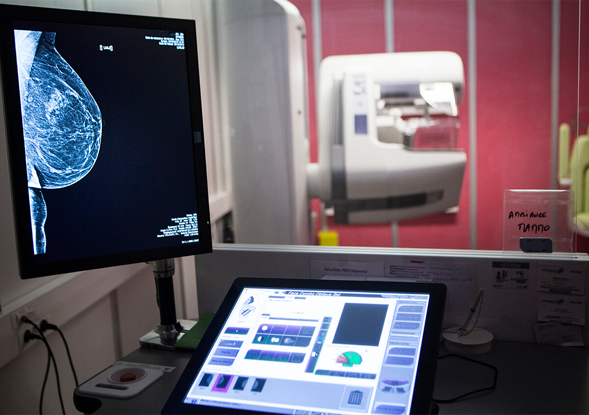
What is a mammogram?
A mammogram takes X-ray images of breast tissue to help diagnose breast cancer and other breast diseases.
Mammograms can often find breast cancer while it’s still very small and in the early stages — even before any lumps or other symptoms are felt. Mammograms are used as a screening test for breast cancer and can evaluate lumps or other abnormalities you or your doctor may have found during an exam.
Types of mammograms
There are two types of mammograms: screening mammograms and diagnostic mammograms.
Screening mammograms are done on a yearly basis when there aren’t any new breast lumps or other new breast symptoms. X-ray images of the breast are typically taken from two different angles during a screening mammogram.
If you’ve found a lump or your doctor is concerned about a specific area on the breast, they may order a diagnostic mammogram. This simply means additional images of the breast will be taken and the radiologist knows to pay special attention to the area of concern.
Your healthcare provider may order a diagnostic mammogram if you’re experiencing new breast symptoms like a new lump, pain, or nipple discharge. A diagnostic mammogram may also be ordered to thoroughly evaluate a specific area of concern on your screening mammogram or if you have a history of breast cancer.
Your radiologist will frequently take additional specialized views of your breast during a diagnostic mammogram, and you may also have a breast ultrasound during your appointment.
Diagnostic mammograms may take longer than a screening mammogram, but you will have your results and recommendations for the next best course of action before you leave our facility.
Diagnostic mammograms are also used if a woman has had a history of breast cancer.
What happens during my mammogram appointment?
After you arrive for your mammogram appointment, you will be asked to change out of your shirt and bra and into a hospital gown.
During the mammogram itself, the breast is compressed between two plates (called compression paddles) to hold your breast in place and capture images. At this time the technologist will ask you to lower your gown to place your breast directly into the mammogram scanner.
Each breast is examined one at a time and while the images are captured you will be asked to hold your breath for a few seconds.
There is no downtime or recovery needed after your appointment and you may return to normal activities.
After your screening mammogram, one of our dedicated radiologists will review the images and report the results to your healthcare team.
At Southern Arizona Radiology Associates, we offer mammograms as well as other types of breast imaging including ultrasounds and breast MRI. You can conveniently schedule your mammogram yourself so you can pick the best time for your schedule.
How old should I be when I get a mammogram?
Annual screening mammography starting at age 40 has been shown to reduce your risk of dying of breast cancer. This is why the American College of Radiology and Society of Breast Imaging recommend regular screening mammograms in women 40-and-older (including 40-49).
While starting screening mammograms at 40 years old is recommended, your healthcare provider may determine you are at a significantly increased risk of breast cancer compared to the general population.
If you have an increased risk, they may recommend starting screening mammograms earlier than 40, and as young as 30 years old.
How often should I have a mammogram?
Once you start having screening mammograms, they are done once a year.
Does a mammogram hurt?
You may feel some discomfort due to the pressure caused by the compression paddles during your mammogram. One way to reduce this discomfort is to schedule your screening mammogram within one week after your menstrual cycle when your breasts are the least tender.
Screening mammograms are also done very quickly meaning any discomfort you feel will only last a few moments.
The technologist completing your mammogram will be able to talk to you the entire time, so you can let them know if you are feeling any discomfort during your procedure.
There are no side effects of mammograms or precautions you need to take afterwards making it a quick and easy way to screen for breast cancer. Many women schedule them before they go to work or on their lunch break because they’re so quick and easy to complete.
3D Mammograms (tomosynthesis) in Tucson and Sierra Vista
Rather than creating two-dimensional images, 3D mammograms (breast tomosynthesis) take several images and create a three-dimensional series of images of the breast using a computer.
3D mammography/tomosynthesis are widely considered the standard of care and have replaced traditional 2D mammograms in most places, including our Tucson and Sierra Vista locations.
How much does a mammogram cost in Arizona?
The price of a mammogram usually depends on if you have insurance and what type of insurance you have. Many insurance companies cover yearly mammograms in full which means you don’t have to pay anything. Sometimes they require a co-pay for your visit.
There are also community and national resources available that can help pay for your mammogram if your insurance does not cover it or you do not have insurance.
When you call our office to schedule your appointment, we will provide you with an estimated payment needed from you at the time of your visit.
Other types of breast imaging
In addition to mammograms, the radiology team at Southern Arizona Radiology Associates can perform several other breast imaging procedures.
Breast Ultrasound
A breast ultrasound uses sound waves instead of X-rays to create images of breast tissue. Ultrasounds are noninvasive and are not painful.
Breast ultrasound is not a replacement for mammography, but can be performed with a mammogram if a particular area of your breast needs a closer look.
There are times when a breast ultrasound may be performed without a mammogram, for example to evaluate problems in younger patients, usually under 30 years old.
During a breast ultrasound, your technologist will apply gel to the breast and scan your breast with a handpiece called a transducer. The images are then reviewed by your radiologist with results and recommendations provided before you leave.
Our radiologists may also use ultrasound technology when performing breast biopsies.
Breast MRI
If someone is considered high risk for breast cancer, they may need a breast MRI in addition to their mammogram to screen for breast cancer. Talking with your healthcare provider to determine your lifetime risk of breast cancer can determine if you need a breast MRI.
Breast MRIs can sometimes detect breast cancer not previously found on a mammogram but should not be used as a replacement for a mammogram. Another reason for a breast MRI is to assist with breast biopsies.
Rather than X-rays, breast MRIs use a powerful magnet to create detailed images of breast tissue. Breast MRIs also require the use of contrast that is administered through an intravenous line placed in your arm.
Like other MRIs, you will be in a confined space during the procedure so talk with your doctor if you are worried about feeling claustrophobic.
Breast MRIs are reviewed by the radiologist and a report is sent to your healthcare provider.
Mammogram Near Me
If you need to schedule your mammogram and live in Southern Arizona — Southern Arizona Radiology Associates is here to help at our convenient Tucson and Sierra Vista locations.
We offer mammograms, breast ultrasounds, breast MRI, and breast biopsy services so you can complete whatever breast imaging you need all in the same place.
You can schedule your own mammogram and other breast imaging services within 24 hours after we receive an order from your healthcare provider. Schedule your mammogram with our radiology team in our Tucson or Sierra Vista locations by calling (520) 335-6849 today.